This August, I attended the Shanghai International Innovation competition and even though it did not have a heath related theme, half of the pitches had a medical application indicating the growing number of healthcare innovation companies.
The winner of the UK final was Eva Diagnostics; founded by former students of Imperial College started with the aim of using technology as a platform to aid healthcare providers to strengthen patient-centered practise by accessing up-to-date patient information. They have developed blood based home monitoring devices with the potential to improve patient quality of life and efficiency measures by promoting the management of chronic conditions and diseases.
Anemipoint is a small handheld device able to accurately measure levels of haematocrit (volume of red blood cells) and haemoglobin (protein within red blood cells) from a drop of blood in 30 seconds. Anaemia affects 3% of men and 8% of women in the UK and is defined as an inadequate amount of red blood cells or haemoglobin leading to a reduced oxygen carrying ability. The device addresses the blood donation, maternity health and nutrition market; with over 100 million blood donations given per year this is a fast way of testing samples, also efficiency in determining whether medication for anaemia treatment is needed by mothers’ is improved. Data is not only used as a diagnostic tool but a secure Cloud based system allows real time data of anaemia prevalence to be collected. AnemiPoint has been tested against the current gold standard and is able to provide the same quality of results. The system has been adapted to work in countries with limited resources; running on a single power charge and requires no reagents, making it sustainable.
HemiStat their other monitor can track symptoms, measure white blood cell count and continuously monitor an individuals response to chemotherapy setting the basis for a more personalised patient healthcare plan in the future. It is the first home monitoring assessment for chemotherapy and is a much needed asset as a reduced white blood cell count is a common side effect and can have dangerous consequences if not handled. Every year 5% of patients develop a serious infection caused by low white blood cell numbers and the device can aid in early detection; helping to better recovery times. It has the potential to save time and money through reducing the number of unneccesary hospital visits as a patients blood count may be too low (previously only able to measure at a hospital) to carry on their next chemotherapy treatment.
The company aims to market launch AnemiPoint by December 2016 and HemiStat in 2018.
After taking a long break from blogging but definitely not reading about science, I have decided to start off the academic year by attending the Royal Society of Medicine’s second innovation summit this year to get my mind in the right headspace.
As always, listening to the speakers truly inspires me and opens my eyes to the vast possibilities of Medicine. Throughout the day I heard from those discussing tackling global health challenges, technology in the applied medical setting and innovative research and I will share with you four of the talks I heard about.
1.
Blood bank shortages is an ongoing problem with around 40% fewer blood donation volunteers than a decade ago in the UK. A new ‘natural’ lab grown source of blood being developed by Dr Joanne Mountford and Professor Marc Turner who were granted the £5 million Strategic award by the Scottish National Blood Transfusion Service could potentially solve this issue.
Current obstacles that need to be overcome with donated blood are; immune incompatibility, sufficiency, transfusion transmitted infection and iron loading (seen in those with β thalassemia form multiple blood transfusions). The researchers use donated skin cells (fibroblasts) which are then genetically reprogrammed forming induced pluripotent cells which are able to self renew and expand indefinitely. By using iPS cells rather than embryonic stem cells collected from unused IVF embryos, it avoids the ethical considerations to think about and the possibility of gene expression. It was also important to them to make sure the blood was ‘natural’ and not made form synthetic materials.
In the lab they are currently able to grow blood with fetal haemoglobin expression (α/γ chain) which is viable in adults and due to their young nature they could give a longer therapeutic benefit and reduce the frequency of transfusions whereas blood bags contain a mixture of cell ages. The cells are matured in suspense cell cultures using growth factors and cytokines with the entire process taking about 30 days.
The aim is to create O Rhd negative blood which can be given to 98% of the population, however challenges they spoke about included overcoming membrane instability after enucleation. It is essential that the nucleated normoblast is converted to a biconcave shape giving them a larger surface area to absorb oxygen. This is checked by marking the proteins with fluoresce to see via laser or stain under a microscope. Furthermore, at the moment cells are only grown to obtain protein and not for the actual cell component so volume reduction and separation technology needs to be developed in order to scale up the RBC production. To meet clinical requirements, 2×10¹² RBC would need to be generated, found in a standard red blood cell concentrate.
They have come a long way since starting this research as they are now able to make 5ml of blood (10% of a unit) efficiently and hope to start the first human trial in late 2016. This concept of natural (non-synthetic) blood looks to be an innovative tool in the future with the ability to reduce NHS costs in disposing of unused donated blood and delivering to developing countries where blood loss during childbirth can save hundreds of thousands of lives.
2.
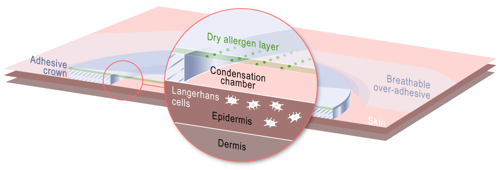
French company DBV Technologies are revolutionising food allergy treatments by developing the Viaskin patch. Current immunotherapy treatment for food allergies (to build up tolerance) are by subcutaneous injections which can lead to itchy and swollen skin. This patch administers the allergen to intact skin avoiding contact with blood increasing tolerability therefore lowering the risk of a systemic, allergic reaction in case of chance exposure.
Viaskin is a non-invasive electrostatic patch that hopes to be a technique to desensitise and improve patient’s tolerability by making the immune system used to the allergen by placing small amounts of the allergen onto intact skin.
The patch consists of a dry allergen layer, a condensation chamber to increase permeability of the skin and solubilises the antigen allowing it to penetrate through the epidermis. Here the antigen is able to reach Langerhan cells which leads to draining of lymph nodes, antigen processing and presentation to T cells which stimulate the immune response and T memory cells. This method uses a passive pathway with no contact with blood to avoid an allergic reaction.
Mechanism of Viaskin patch
Preliminary tests done by DBV show that epicutaneous immunotherapy (EPIT) may induce an epigenetic change (specifically a modification of DNA methylation).
Dr. Pierre-Henri Benhamou, Chairman and CEO of DBV Technologies has previously said that, “EPIT appears to be the only method of specific immunotherapy able to confer durable protection in case of accidental exposure to allergens. We believe that this is probably due to the ability to influence the immune response to allergens at the genomic level”.
A 2014 study published in the British Society for Allergy and Clinical Immunuology concluded that regulatory T cells (Tregs) mediated mechanism of EPIT resulted in long-term protection against eosinophilic disorders* in peanut-sensitised mice which is a very positive outcome.
As it is easy to use and safe, early intervention in children could be a realistic possibility without the risk of anaphylaxis could prevent the disease developing. With over 5 million peanut allergy sufferers in Europe and the USA, if successful could make a huge difference to their lives. DBV Technologies are also currently working on using Viaskin technology to treat milk and egg allergies and an alternative to some vaccines.
*Too many eosinophils (WBCs) produced causing chronic inflammation and tissue damage.
3.
All the way from Michigan, Glenn Green and Scott Hollister came to speak about just the second procedure of it’s kind to use a custom biodegradable implant. The 3 year old patient’s condition (18 months old at the time) had left him hospital bed-ridden with ventilator levels at their maximum pressure and was even placed in a medically-induced coma to keep him alive. The condition the child had is called, Tetralogy of Fallot with absent pulmonary valve syndrome, only 3% of those with Tetralogy of Fallot also missing the pulmonary valve.
Due to the absence of the pulmonary valve, pulmonary regurgitation occurs (retrograde flow of blood from pulmonary artery to the right ventricle). This lead to him developing severe tracheobronchomalacia, a disease that weakens the muscles around the trachea. It was so debilitating that he would turn blue sometimes four to five times a day as the smallest movement could cause his airways to collapse to the point where they were just small slits. There are approximately 1 in 2,200 babies born with tracheobronchomalacia but severe cases are rare.
In December 2013, his condition was worsening and plans needed to be finalised with emergency clearance by the FDA to create and implant a bioresorbable tracheal splint made from a biopolymer. After taking a CT scan of the child’s trachea and bronchi, the splint was produced using a system that takes an image computer model alongside 3D laser printing to create a custom-design. They had about 20 to 30 different models as they knew how it looked on screen but were prepared for any contingencies.
In 2014 Green assisted Dr. Richard Ohye in performing the surgery, two splints were sown around his left and right bronchi to allow expansion of the airways and support to aid growth. Since the operation, the boys airways have remained open and ventilation levels are less than a quarter of what they used to be. It shall take around three years for the trachea to remodel itself and that is about how long it will take for the splint to be reabsorbed.
4.
In May 2015, surgical teams from Houston Methodist hospital and the University of Texas MD Anderson Cancer centre collaborated to perform the first composite vascularised allo-transportation surgery alongside craniofacial tissue transplant.
A 55 year old man presented with a failing kidney and pancreas from a previous transplant in 1992 as a result of Type 1 diabetes and a previous history of leiomyosarcoma (rare cancer of soft muscle tissue) which left him with a large, deep wound on his scalp and exposure of the skull bone. Due to the risk of infection from his head wound, surgeons were hesitant to perform a new organ transplant, however Dr. Jesse Selber saw an opportunity here. As the patient was already immunosuppressed from previous solid organ transplants, the high threshold that ordinary transplant candidates would have to meet due to increased risk of taking long-term immunosuppressants would not be a problem. Carrying out a skull and scalp transplant simultaneous to the double-organ transplant would mean the anti-rejection medication could support them all.
A wait of 18 months ensued to find the correct donor as he already had other antibodies in his blood from the previous transplant and many donors could not be used. Selber’s skills in microvascular technique were essential to this type of surgery as blood vessels 1/16th of an inch need to be connected to the scalp quickly to restore circulation. Transplants involving tissue structures such as skin, bone, blood vessels and nerves such as this one are called vascularised composite allograft and techniques which are still relatively new and techniques are still progressing. Dr Selber expressed to us that while this surgery took many months of meticulous planning and organising, they had to improvise after realising that the donor’s skull was much thicker compared to the remainder of the patient’s and had to reshape the skull with saws and drills in order to make it fit. Being the first surgery of it’s kind, the surgeons were amazed when seeing the blood flow through the scalp once it had been connected as the grey organ was brought to life. The pancreas and kidneys were then transplanted with surgeons having to work around the calcifications formed from his previous failing organs. After 15 hours, the four transplants were complete and the patient continues to be closely monitored using Skype consultations as a method of keeping up to date so no travel time is needed unless in emergency.
Dr. Jesse Selber also spoke about how he has now asked the patient to take daily photographs of his head to oversee the skull and scalp transplant healing to get a good overview. Selber recalls his fascination at watching the patient’s scalp sweat the Wednesday after the surgery in a hot recovery room and how it was unexpected due to the nature of not knowing what is to come as it is a new type of surgery. The doctors expect the patient to have rejection episodes, as is common with allografts. In fact just a few weeks ago there was a scare that the body might be rejecting the scalp as a large rash was appearing on his scalp however after testing and a revising of medication there are no signs of rejection. This surgery was innovative and very complex with around 50 health professionals all gathered in one surgical theatre to make this possible.
Hope this has given you a glimpse and want to go out and innovate!
I had the great pleasure of attending the RSM’s 10th Medical Innovations summit again this year and thought I would share a few of the wonderful presentations I saw.
1. Dr Geraldine Hamilton having been frustrated by the lengthy time for clinical trials to take place and have a new medicine approved on the market was inspired to create ‘Organs-on-Chips’.
Simply put it is a polymer about the size of a USB stick with three fluidic channels. The centre is a porous, flexible membrane where human cells can be added above and below and vacuum channels on the sides. In order to make it realistic the cells can be stretched and contract to experience the same mechanical forces as they do in the body. Air channels are above the cells and a liquid containing nutrients is passed through the blood channel underneath.
Using microchip computer technology, these devices produce levels of tissue and organ performance not possible with typical culture systems. A use of the technology is to mimic infection this was shown to us on a video where lung cells had bacteria infecting them, leukocytes were then added and we saw them engulfing the bacteria onscreen. ‘Organs-on-chips’ allow scientists to investigate the biochemical, genetic and metabolic responses of the cells.
The end goal is to connect different ‘organs-on-chips’ via fluid to create a ‘human-on-chip’ and recreate sufficient functionality to better predict the outcome in humans. Later on, there could be the possibility to put your own stem cells on the chip and therefore be able to see a personalised reaction to new medication. This technique could really revolutionise the way we test efficiacy of drugs via clinical trials and potentially eradicate the dependency of animal testing.
2. Neeti Kailas made a speech about designing a hearing screening test device that could be used under the busy constraints of Indian paediatric clinics. The company, Sohum Innovation Lab, aims to create “market-driven solutions to improve the health and incomes of people living in resource-poor settings.”
This was of particular interest to me having carried out work experience in September at a paediatric audiology clinic in East London where I was able to witness the different hearing tests being performed. With the Healthy Child programme in the UK it is routine to perform an otoacoustic emission test on newborns and sometimes a secondary automated auditory brainstem response test (which is what the Sohum device uses).
On the other hand, with 28 million babies being born each year and no routine screening in India, many cases of hearing impairment are discovered too late and can have a detrimental impact on the child’s later development. When accompanying a friend in India to a paediatric clinic she saw all too clearly that the healthcare system needed to be changed. When designing the hearing test device she needed to overcome some difficulties specific to clinics in India, such as the noisy environment, inability to afford soundproof rooms, need for quick tests in order to deal with busy crowds and can be used by unskilled workers. As a designer, she used her skills to create a non-invasive portable device that is self-explanatory and can be performed in three minutes. The device has the backing of the Department of biotechnology in the Indian government and they hope it may lead to hearing tests being routinely carried out.
3. Dr Claire Guest is the founder of Medical detection dogs, where they train dogs to help manage human sufferers of certain diseases. For example, people who have diabetes can be signalled by their dog when they are about to have a hypoglycemic attack. This is life-saving training as there are more than 300,000 people living in the UK with type 1 diabetes and it costs the NHS £1,700 per hypoglycemic episode. Dogs have 125- 300 million nose receptors and are therefore far more superior in smelling biochemicals produced in humans.
The other side of the charity is spent training dogs to detect cancer. The idea of dogs sniffing cancer first came about in 2004 when a paper was published in the BMJ about investigating bladder cancer via dogs smelling samples. In 20 minute intervals, the dogs are individually led round a carousel containing samples (in breast cancer this would be a breath sample on a fibre filter). The dogs are led round the carousel and taught to stop and sit when they detect a cancerous sample. It costs £10,000 to train a dog for 4 weeks and each dog is only trained to detect one type of cancer so there is no confusion for the dog. All samples used are before the patient has undergone any treatment for the cancer so that there is less chance of the dogs sensing the chemicals from chemotherapy.
The potential is that samples could be sent for the dogs to test and aid consultant’s in deciding whether to go ahead with an invasive procedure (e.g. biopsy), providing a second line screening. Cancer detection dogs can also be used to help scientists research into E-noses and enhance early detection via cheap tests. So far there have been promising results with a recent study in Italy showing sensitivity 98-100% and specificity 98-99% in recognising specific volatile compounds in prostate cancer samples. The number of false positives was significantly lower than traditional PSA tests which have a 75% rate.
4. Motivated by his grandfather’s struggle with Alzheimer’s and his family’s subsequent worrying about his frequent wanderings Kenneth Shinozuka has gone on to develop a wearable sensor on the foot to alert carers via an app when the patient steps onto the ground and is about to wander. There are currently 5.2 million sufferers of Alzheimer’s disease in the US with 65% of them wandering with a $220 billion cost in paying caregivers.
Kenneth first identified three main obstacles to overcome in order to make his vision into a reality; design a sensor, create a circuit and develop a phone app. He decided to use a film sensor with electrically conductive, pressure sensitive ink particles. The circuit uses Bluetooth low energy technology which can be driven by a coin-sized battery so it is not bulky. The device works because when pressure is applied the connectivity in the particles increases and electrical resistance can be measured.
Two prototypes have been made; one being integrated into a sock and another which is re-attachable. The device has since had an 10% success rate in detecting all his grandfather’s 437 cases of wandering. Kenneth is now working on developing the sensor to be able to track the patient using geo tagging and also commercialising the product.
I hope these snippets of some of the talks I heard have given you some inspiration to develop your own medical technologies so we can keep on advancing in healthcare. My next blog post is going to be a special feature on another speaker from the RSM summit, Dr Jonathan Landy who is the co-founder of Figure1 and it will feature a Q&A so look out for that.
With an ageing population, neurodegenerative diseases are becoming more prevalent and an effective solution needs to be found fast. The Alzheimer’s Association (AA) in a 2012 report estimated that a new case of will appear every 33 seconds by 2050. In neurodegenerative disorders such as Alzheimer’s and Parkinson’s disease there is a loss in the number of synapses which leads to symptoms such as memory loss.
Researchers at the University of Leicester and University of Cambridge recently published a study in Nature looking at the effects of hibernation on brain synapses on mice. It was found that the lost synapses when going into hibernation were recovered when coming out of it. They next wanted to investigate if the level of protein RBM3 (a protein which is still produced despite cold temperatures) played a role in the regeneration of synapse.
Brains of mice from three groups (normal, Alzheimer’s disease sufferer and prion disease sufferer) were studied during cooling to 16-18C and rewarming (although some were not cooled and had their levels of RBM3 chemically altered as a control), comparing the number of synapses and level of RBM3.
It was found they had increased levels of RBM3 during cooling and it remained elevated for up to three days after. The mice which had been cooled survived for an average of seven days longer which implies cooling gives an added protection. However, those with a more advanced version of the diseases lost synapses when cooled and could not regrow them. In addition, it was found that disease progression sped up when RBM3 levels were reduced.
It was concluded that protein RBM3 is involved in the pathway of synapse regeneration in mice. The research showed how cooling is protective against the loss of synapses in the early stages of rodent forms of Alzheimer’s disease and prion disease but in advanced stages it was suggested that cooling may not be protective due to no increase in the level of RBM3s. It has already been found from other investigations that therapeutic hypothermia leads to increases in RBM3 and if this pathway could be stimulated in humans it may lead to a potential treatment for neurodegenerative disorders but this is at a very early stage.
Last Saturday was the RSM’s Annual Medical Innovations Summit and I luckily managed to get a booking for the afternoon session. I will just share with you some of the amazing innovations presented.
A key theme throughout the day were apps created by healthcare services (e.g. GP practices) in order to allow better communication with their patients, improving access to primary care services. The MyGP app in association with Brook Green Medical Centre allowed patients easy monitoring of chronic health problems, access to their medical history, appointment calendar and the ability to find local healthcare services.
A concern voiced by a member of the audience was about confidentiality and how this would be addressed in case the phone was stolen or hacked into. The GP explained that while this is a possibility, the technology allows for it to be shut down once reported and that there is probably a higher likelihood of valuable information from paper files (as the system currently is) being stolen from a briefcase or on a ward rather than through the app.
LifeSaver water bottle was designed by Michael Pritchard after he saw that water aid given in disasters was slow and inefficient. It works simply by having a cartridge in the middle of the bottle which has pores 15 nanometres wide so that bacteria, viruses and waterborne pathogens are trapped within. The pump on the lid is operated manually and forces the water through the filter to release safe drinking water.
He even showed us that it worked by pouring in murky water filled with nasty materials (I shall spare you the details) into the bottle and then drinking it afterwards!
Owlstone presented a microchip chemical sensor that was placed in a breathalyser in order to detect chemical compounds (via mass spec or gas chromatography) found in people with certain medical conditions such as cancer, TB or diabetes. The idea of detecting diseases by chemicals in the breath is not new and Hippocrates was said to have smelt the breath of his patients as a way of diagnosis. The chemical breathalyser is an exciting new way of giving early diagnosis and may be seen in the near future. The sensor technology can also be used in bodily fluids to detect illnesses like Inflammatory bowel disease. This technology will be very helpful as it gives quick results, can be used instead of invasive procedures (e.g. colonoscopy) and at a lower cost.
A study funded by the National Institute of Neurological Disorders and Stroke (NINDS) discovered that the brain may clear toxins that have built up during the day. We already know that sleep is fundamental in safeguarding memories and now it seems that the brain may increase the space between brain cells allowing the fluid to flow through rapidly. This is part of the glymphatic system which controls the flow of cerebrospinal fluid through the brain.
Researchers first injected dye into the CSF of mice to see how it flowed through their brains and monitored their electrical brain activity. They compared the flow rate of when they were unconscious to when they were awake and the later was significantly slower. It was then theorised that the distance between brain cells changed between unconscious and conscious states. This was tested by inserting electrodes into the brain of the mice to measure the space between the brain cells. It was found that the space increased by 60% when the mice were unconscious, allowing CSF to flow more freely.
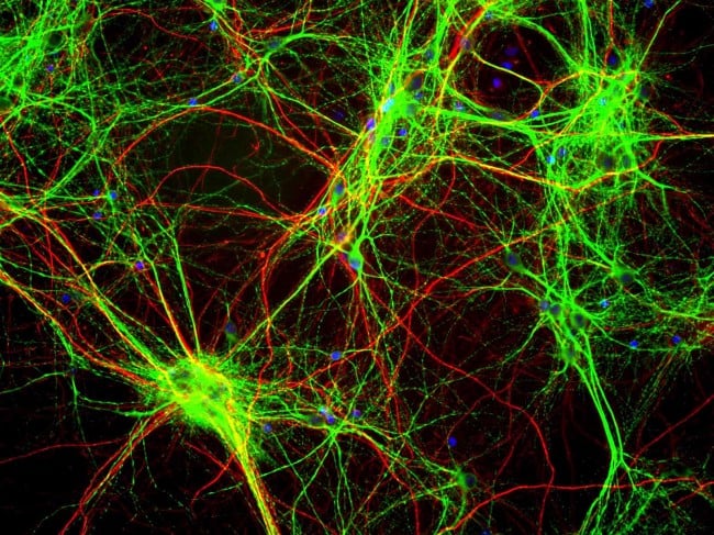
Stained rat brain cells
Brain cells called glia control the glymphatic system by shrinking or swelling. The hormone noradrenaline is known to control cell volume and was discovered to be less active during sleep. This was found by giving the mice drugs which blocked noradrenaline, this subsequently induced unconsciousness. The hormone is usually released when the brain needs to become more alert, for example, in response to fear.
Other studies have proposed that toxic molecules collect in the space between brain cells. When the protein beta-amyloid (associated with Alzheimer’s disease) was injected into mice, it was shown that there was an accumulation of the protein when they were sleep deprived. This suggests that the glymphatic system is key in clearing the toxic molecules.
The study carried out may be very helpful in understanding the clearing of toxins and that the glymphatic system may need to be targeted in order to treat certain neurological diseases such as Alzheimer’s.
Note: The research was carried out at the Nedergaard Lab, University of Rochester Medical Center and was published in Science.
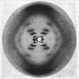
Over the summer I visited the Photo 51 exhibition at Somerset House in association with King’s College London. It was there to celebrate the 60th anniversary of Photo 51 which was crucial in the discovery of the structure of DNA. Photo 51 is an iconic image in scientific history, it is the X-ray diffraction image of DNA which was taken in May 1952 in the lab of Rosalind Franklin by her PhD student Raymond Gosling.
X-ray crystallography was used to produce images by sealing a small sample of hydrated DNA in a camera, in front of a piece of x-ray film. An X-ray beam then shone at it for more than 60 hours. The beam of X-rays scatter and produce an image from which a 3D structure can be determined. In the lab, they had been investigating whether the humidity at which they kept the samples would affect the images. Photo 51 was the image taken at the highest humidity circa. 92%.
The darker patches in the picture show the DNA’s bases which make up the genetic code. The diffraction pattern (cross shape) shows the helical nature of the strands as the arms of the cross display planes of symmetry. Photo 51 helped Watson and Crick with calculations on size and structure to develop the model of DNA.
With the United Nations Environment Programme, having recommended a shift to a vegan diet, recent torrent of meat scandals on the news headlines and a rise in number of celebrity vegans, could veganism be soon turning mainstream? Today, among 2.5% of the population in the USA identify themselves as vegans and according to Google trends, this shift is on the increase with more people adapting to a vegan lifestyle. So, what is so appealing about this way of living?
The short answer is this: high levels of saturated fatty acids and cholesterol are generally found in meat diets which are major contributors to increased risk in cancers, coronary heart disease, stroke and diabetes whereas in direct contrast, the high quantity of non-nutritive phytochemicals including carotenoids, all have demonstrated to be beneficial in enhancing cell function and protective against cancer, are naturally present in vegan diets. The high levels of fibre contributes to bowel health and to lower cholesterol levels as LDL receptors are promoted. Whilst an absence of saturated fat from meat benefits the heart in addition to the many other phytochemicals beneficial properties such as antioxidant, antineoplastic (checking maturation and proliferation of malignant cells), anti-inflammatory and/or anti-carcinogenic (reduces the occurrence of cancers) properties.
Whilst high levels of contention exist between authors on the many disparate study results on biochemical effects of varying dietary concentrations, there is evidence pointing to a link in increased breast and colon cancers risk to the excessive intake of essential amino acids found in meat diets that increase the Insulin-like growth factor 1 (IGF-I) activity. With its naturally reduced dietary protein of less essential amino acids, low-fat vegan diets are shown (in animal studies with soy protein) to correlate with reduction in the IGF-I activity. Vegan diets seem to be a better answer than meat diets to reducing the risk of developing cancers including prostate cancer risk associated with high IGF-I activity in men due to higher insulin resistance. All other factors rank equally, with more favourable lipid profiles, lower BMI, lower systolic and diastolic blood pressures associated with vegan diets than meat diets, the former is the clear winner in reducing coronary heart disease.
Though having lesser essential amino acids than meat proteins, many plant proteins contain a higher amount of non-essential amino acids than most animal food products, which are beneficial in helping to promote the secretion of glucagon, an essential part of the feedback system together with insulin, that keeps blood glucose at stable levels. In respect to diabetic risk, again, low-fat vegan nutrition along with exercise, helps more effectively than the meat diets in diabetic control and to lower blood pressure.
Of course, there are some downsides to a vegan diet such as if substitute nutrients are not taken, there could be a risk of nutritional deficiencies by eliminating animal products. Particular nutrients include vitamins B-12, D, calcium, and omega-3 fatty acids. Studies concluded that vegans have lower protection against hemorrhagic strokes due to a reduction in IGF-I activity, therefore decreasing the structural integrity of cerebral arteries. A reduced growth factor in vegans could be responsible for the increased risk. However, by reducing salt intake and taking other realistic precautions to encourage cerebrovascular health this problem can be faced head-on.
Becoming a vegan is not a light-hearted decision but it could have serious benefits. It’s always best to do some more research and decide for yourself!
Sources:
Vegan proteins may reduce risk of cancer, obesity, and cardiovascular disease by promoting increased glucagon activity. (Med Hypotheses. 1999)
IGF-I activity may be a key determinant of stroke risk–a cautionary lesson for vegans. (Med Hypotheses. 2003)
A scientific review of the reported effects of vegan nutrition on the occurrence and prevalence of cancer and cardiovascular disease